From CLARITY Wiki
(→Sandwich sample with second glass dish) |
(→Sandwich sample with second glass dish) |
||
| Line 36: | Line 36: | ||
File: Pressing_down_to_seal.jpg|Pressing down to seal |
File: Pressing_down_to_seal.jpg|Pressing down to seal |
||
File: Tissue_sample_between_two_dishes.jpg|Tissue sample between two dishes |
File: Tissue_sample_between_two_dishes.jpg|Tissue sample between two dishes |
||
| − | + | File: Side_view_of_parallel_glass_dishes.jpg|Side view of parallel glass dishes |
|
</gallery> |
</gallery> |
||
<br clear=all> |
<br clear=all> |
||
Revision as of 06:48, 16 April 2014
Before mounting the sample for imaging, incubate the sample in the mounting solution for several hours or overnight (until the sample is visually transparent. Suggested methods for mounting large tissue samples (whole brain) and smaller sections (<1 mm thick brain slice) using the imaging chamber supplies are detailed below.
Contents |
Whole brain mounting for confocal imaging
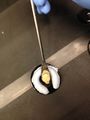
Form a putty cylinder
Roll a piece of BluTack putty into a cylinder with uniform thickness. The cylinder should be several centimeters in length with a thickness greater than that of the brain sample.
Rolling putty into a cylinder
Putty cylinder


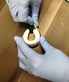
Place putty on a glass dish
Shape the putty cylinder into a horseshoe U shape. Place the putty onto the flat side (outside) of a Willco glass dish. Using a pipette tip, press the outside edge of the putty U down onto the glass dish to help seal it.
Shaping putty cylinder
Horseshoe shaping
Putty on glass dish
Side view of putty on glass dish
Sealing with pipette tip


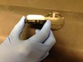
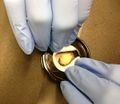
Place sample onto glass dish
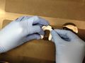
Using a spatula, carefully place the tissue sample on the center of the dish surrounded by, but not touching, the putty. (A mouse brain before clearing is shown for the purpose of demonstration.)
Placing tissue sample on the glass dish
Tissue sample on glass dish


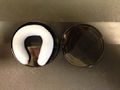
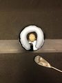
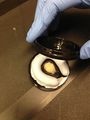
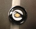
Sandwich sample with second glass dish
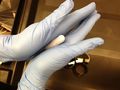
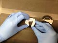
Carefully place the flat glass side of a second Willco dish on top of the putty. Slowly press down evenly on the glass dish, keeping fingers over the putty perimeter, until the glass contacts the top of the tissue sample. Check that both glass dishes are parallel to each other.
Adding second glass dish
Pressing down to seal
Tissue sample between two dishes
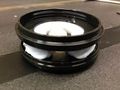
Side view of parallel glass dishes


