Sample Mounting
From CLARITY Wiki
(Difference between revisions)
(→Shape putty into a U on a glass dish) |
(→Place putty on a glass dish) |
||
| Line 14: | Line 14: | ||
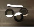
Shape the putty cylinder into a horseshoe U shape. Place the putty onto the flat side (outside) of a Willco glass dish. |
Shape the putty cylinder into a horseshoe U shape. Place the putty onto the flat side (outside) of a Willco glass dish. |
||
<gallery> |
<gallery> |
||
| − | + | File: Shaping_putty_1.jpg|Shaping putty cylinder |
|
| − | + | File: |
|
| − | + | File: |
|
| + | File: |
||
</gallery> |
</gallery> |
||
<br clear=all> |
<br clear=all> |
||
Revision as of 06:26, 16 April 2014
Before mounting the sample for imaging, incubate the sample in the mounting solution for several hours or overnight (until the sample is visually transparent. Suggested methods for mounting large tissue samples (whole brain) and smaller sections (<1 mm thick brain slice) using the imaging chamber supplies are detailed below.
Contents |
Whole brain mounting for confocal imaging
Form a putty cylinder
Roll a piece of BluTack putty into a cylinder with uniform thickness. The cylinder should be several centimeters in length with a thickness greater than that of the brain sample.
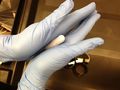
Rolling putty into a cylinder
Putty cylinder


Place putty on a glass dish
Shape the putty cylinder into a horseshoe U shape. Place the putty onto the flat side (outside) of a Willco glass dish.
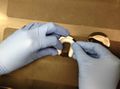
Shaping putty cylinder
